The Eye
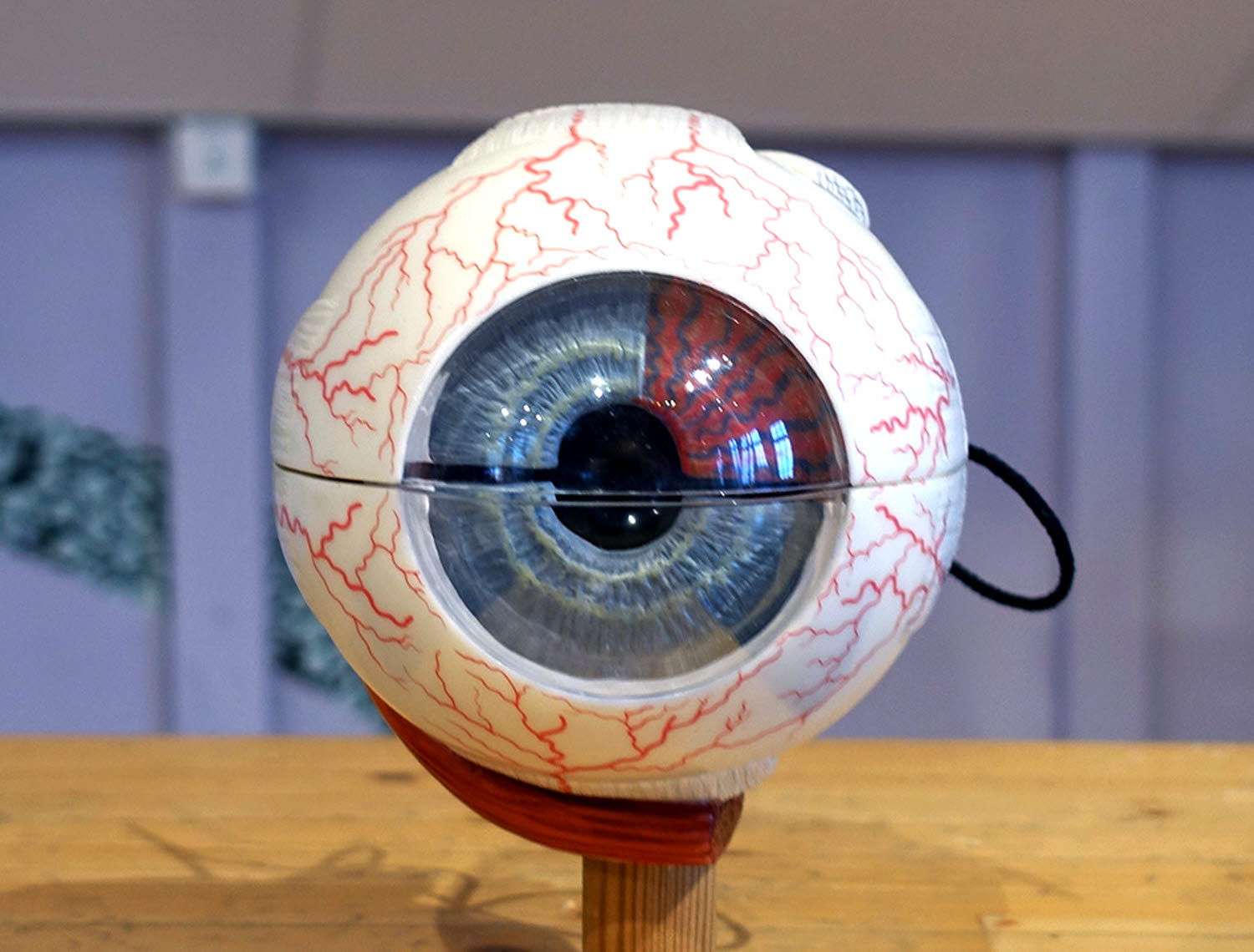
Our eyes enable us to see the world, but how do they do that? Take the model apart to see the tissues that are involved.
Light enters the eye via the pupil. The light is then bent (refracted) in the cornea, the lens and the vitreous body. When the light is refracted, an upside-down image is created on the retina in the rear part of the eye. Electrical signals from the retina are transmitted via the optic nerve to the brain's vision centre in the occipital lobe.
Cells that allow us to see are known as optical cells or visual receptors. They are affected by the information we receive from light. Visual cues are interpreted by the brain and we see three-dimensional images.
The optic cells convert the information into electrical signals, which are redistributed as nerve impulses in the nerve fibres. When the information reaches the brain, you become aware of it. Thanks to this information, we can see.
The eye can only perceive light in certain wavelengths, what we call visible light. Light can have different wavelengths, which we perceive as different colours. Six small muscles help the eye move. The muscles contract to allow us to focus.
The eyeball looks like a flattened orb, and is located in the eye socket. The eyeball is protected by fatty tissue. The eyeball is hollow and contains a gel-like fluid called the vitreous body. The wall of the eyeball consists of several layers. Each layer in the different parts of the eyeball varies in thickness.
We find the rods and cones in the retina. The rods help us see in the dark, but do not distinguish between colours. The cones allow us to see different colours and are used when lighting is good. There are three different types of cone, and they are most sensitive to blue, green and red.
The rods and cones in the retina are in contact with nerve cells, which are also located in the retina. The nerve cells have long projections that together form the optic nerve, which leads from the rear of the eyeball. In the area where the optic nerve passes out of the retina, there is an area without rods and cones known as the blind spot.
The cornea on the front of the eye is completely transparent, so that light can penetrate the eye. The iris has a round aperture known as the pupil. The iris controls the amount of light allowed to enter the eye by using small muscles to control the size of the pupil. You can have a closer look at your own pupils in the Pupil Mirror!
The eye is built like a camera, where the aperture is the pupil, the lenses are the cornea, lens and vitreous body, and the film is the retina. The eye is protected in the eye socket by the eyelid. The eyelid closes the eye and prevents the cornea from drying out. The tear glands produce tears in the upper and outer part of the eye socket. Tears rinse the eye when you blink and keep the cornea free of particles in the air. Tear also protect the cornea from drying out and from bacteria. The tears are captured by the tear ducts in the corner of the eye and flow into the nose.


