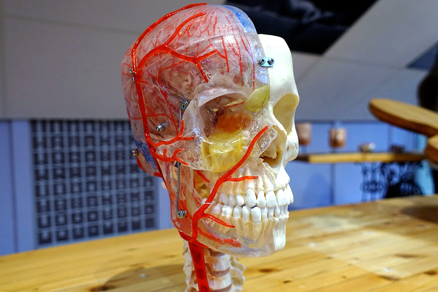
Brain head neuro anatomical model ical
This model shows the vertebrae of the neck and the eight areas of the brain. It also shows the network of nerves and arteries.
The cerebrum
The cerebrum is the largest part of the brain, and the part that is the most developed. The complex functions of the cerebrum make us different from animals. It is divided into two halves called hemispheres. Often, the left hemisphere is the most dominant and governs speech and language. The right hemisphere is more artistic and creative.
The cerebrum looks like a slippery walnut and is filled with winding furrows. Where the furrows are most defined, the brain is divided into different lobes with different functions:
- The frontal lobe: Controls voluntary muscle movements.
- The parietal lobe: Registers tastes and sensations.
- The temporal lobe: Hearing and smell centre.
- The occipital lobe: Vision centre.
Each body part has a special place in both the movement and sensory centres. The most active body parts such as the hands, mouth, lips and tongue take up the most space.
In the outermost layer of the cerebrum contains the cerebral cortex. This is where the cells of the nerve cells live and where our consciousness, thoughts, feelings and memories are formed alongside voluntary and involuntary movements. The cerebral cortex is made of grey matter.
The remainder of the cerebrum is made of white matter, where nerve tracts with grey substances can communicate with each other. This results in interaction between all parts of the brain, not just in the cerebrum.
The cerebrum contains four cavities filled with fluid. These are called ventricles and contain cerebrospinal fluid (CSF). CSF works as a protector and shock absorber for the entire central nervous system – the brain and the spinal cord. The fluid is created in blood vessels in the ventricles and transports nutrients to the brain and removes waste.
The cerebellum
The cerebellum is located beneath the occipital lobe is called the "tree of life" because of its appearance when in cross section. The cerebellum also contains two hemispheres. It is folded just like the cerebrum, but the folds are thinner. In the cerebellum, nerve cells communicate via grey matter between the white matter.
The cerebellum controls our balance and fine motor skills using information from the cerebrum. Information about a body’s position and how we want to move.
The meninges
There are three different types of meninges inside the skull:
- The dura mater that separates the cerebrum and cerebellum. It also protects the entire cerebellum. The dura mater has two layers that are connected by large veins in some places.
- The arachnoid mater is found inside the dura mater. It is web-like and connects to the pia mater. CSF lies between the arachnoid mater and the pia mater. This is where the large arteries that supply the brain with blood can be found.
- The pia mater is closest to the brain and surrounds the brain’s blood vessels.


