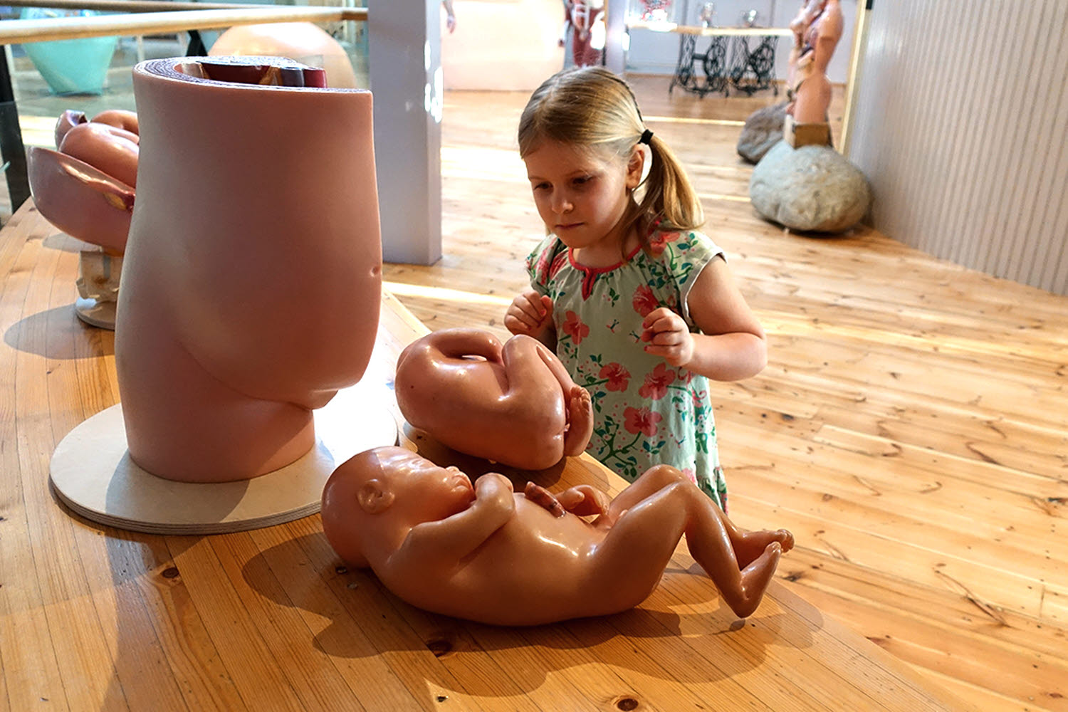
From egg to newborn
Study how the foetus develops right up until birth. The models are partially "hands on", and can be investigated together.
Embryo at one month
The early stages of the placenta can be seen on the model of a month-old embryo. The placenta is where the foetus’s blood obtains its nutrition and oxygen from the mother’s blood.
The placenta grows to meet the oxygen and nutritional needs of a foetus. In the embryo’s first month, cells rapidly divide as various cell types for different organs develop. You can also clearly see the beginnings of the spine and brain. Most organs form later on in the sixth week following conception. The embryo now resembles a human and is called the foetus. The foetus floats in liquid inside the amniotic sac and is protected from impact and pressure.
Third and fourth months
The foetus can now use its hands to grip and its feet to kick.
The fourth and fifth month of pregnancy
Hearing has developed so the foetus can hear. Around this time, the woman will be able to feel the foetus move.
Twins
The twins in the model are fraternal (dizygotic) twins. They developed from two fertilised eggs. Each twin has its own placenta and amniotic sac. Fraternal twins are no different to regular siblings.
The sixth month of pregnancy
At this point, the foetus’s organ system is capable of functioning. The foetus continues to grow in size right up until childbirth.
Childbirth
A normal pregnancy is usually 284 days calculated from the first day of the last menstrual period.
In a normal pregnancy, when it is time for the child to be born, the uterus will have grown from seven to 30 cm and increased in weight from 60 to 1,100 g. The uterus will also contain around one litre of amniotic fluid. The combined weight – including the foetus – is around five or six kilos.
The muscles of the uterus stretch as the foetus grows. This leads to an increase of spontaneous muscle contractions towards the end of the pregnancy. The contractions make the belly tight, like a ball. This is the uterus’s way of preparing for the exertion needed to push out the foetus. We do not know exactly what causes childbirth, but there is probably a link between hormone levels and foetal development.
The opening phase
The opening phase takes around 12 to 18 hours for a person giving birth for the first time. It is often shorter for someone giving birth for a second time. During the opening phase, the uterus slowly expands. Contractions push the baby’s head (or bottom if it is a breech birth) towards the cervix. The head acts as a wedge and dilates (expands) the cervix. The bones of the pelvis increase in girth as ligaments stretch.
The expulsion phase
When the cervix is sufficiently dilated, the mother can help push out the baby. In addition to the uterus’s contractions, the mother can use her abdominal muscles and diaphragm to push the baby’s head into position between the cervix and the vagina. This stage can last from 10 minutes to several hours and is difficult for the mother and most likely for the baby. Once the head has passed the narrowest part of the birth canal, things start to get easier.
The final phase
A few minutes after the baby has been born, the placenta and the foetal membrane is expelled. This is called the afterbirth. The midwife checks that the placenta is intact and nothing remains inside to cause an infection. Muscle contractions in the uterus shut off the mother’s blood vessels so blood loss is as low as possible.
It takes around one month following the birth before the uterus has healed. You can tell from the discharge from the uterus following childbirth. The uterus continues to contract and will finally return to the size it was before pregnancy – a small pear.
Do you want to experience what it’s like to be seven months pregnant? Try the experiment Pregnancy suit!


